D-R. Chebat1, J-K. Chen2, A. Ptito2, R. Kupers3, F. Schneider1,4 and M. Ptito1,4.
1Chaire de recherche Harland Sanders, Ecole d’Optométrie, Université de Montréal, Qc, Canada; 2Neuropsychology, Montreal Neurological Institute, McGill University, Qc, Canada; 3Pet center, Riggs Hospital, Copenhagen, Denmark; 4Danish Research Centre for MR, Copenhagen University Hospital, Hvidovre, Copenhagen, Denmark.
1Chaire de recherche Harland Sanders, Ecole d’Optométrie, Université de Montréal, Qc, Canada; 2Neuropsychology, Montreal Neurological Institute, McGill University, Qc, Canada; 3Pet center, Riggs Hospital, Copenhagen, Denmark; 4Danish Research Centre for MR, Copenhagen University Hospital, Hvidovre, Copenhagen, Denmark.
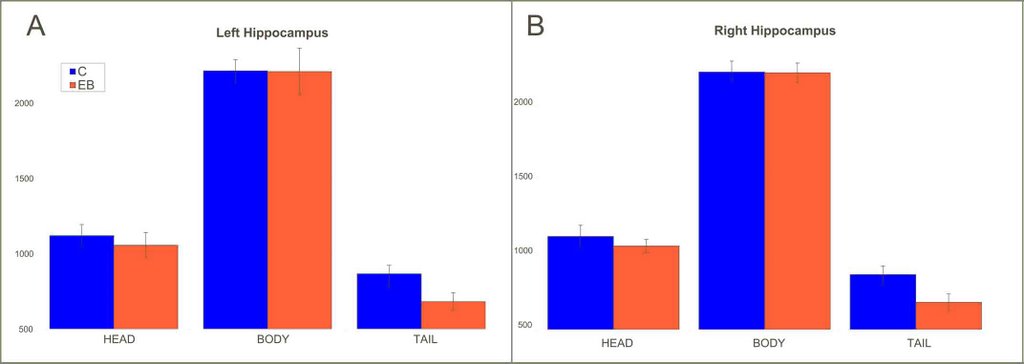
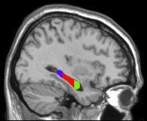
There is a relation between the hippocampus and coding of allocentric space(Crane & Milner, 2005). Since navigation is a highly visual task, what structural changes could be associated with non-visual navigation? This study compares hippocampal volumes of 10 early blind, and 10 sighted controls. Structural MRIs were obtained with a 1.5 T Siemens and hippocampi were manually segmented in three parts: anterior, body and posterior. Mapping was done by two independent experimenters blind to the conditions. MR scans and hippocampal mappings were also normalized into stereotaxic space (MNI). Hippocampal delimitations were binarized and the reccurence of each voxel as belonging or not to the hippocampus was compared between groups (Chi2). No differences in total hippocampal volumes were found and there was no within group differences for left versus right hippocampal volumes. Early blind subjects had significantly smaller posterior hippocampi compared to controls (ANOVA, p<0.05).
Supported by the Harland Sanders Chair in Visual Sciences and the Danish Medical research Council.


No comments:
Post a Comment